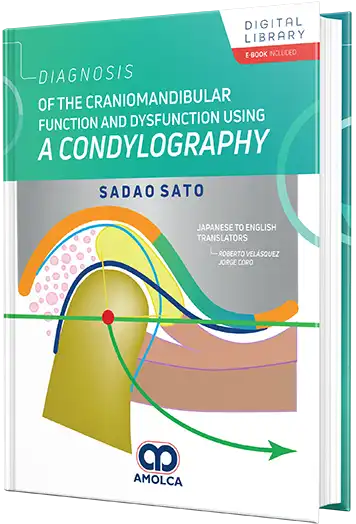
Diagnosis of the craniomandibular function and dysfunction using a condylography (edición en Ingles)
Oclusión
Ebook
200,00 USD ($)
Descripción
Ficha Técnica
9786287681507
Lujo gofrado
Dura
206
2024
0
1.34 kg
1
Section I Theory / 1
Chapter 1. History of Occlusion and Mandibular Movement Research
1) The dawn of mandibular movement research
2) Gysi’s Axis Theory
3) A new era of mandibular movement research
4) The age of Gnathology in California
5) The paradigm shifts in the field of gnathology
6) A new era of gnathology
Chapter 2. Structure and Function of the Cranio-mandibular System
1) Evolution of morphological changes in the human temporomandibular joint
2) Cranio-mandibular System (CMS)
Chapter 3. Concept of Mandibular Position - Clinically Required Mandibular Position
1) Definition of Centric Relation
2) Definition of Mandibular Position
3) In search of physiologic treatment, mandibular position
4) Misconceptions about the hinge axis and mandibular position
5) The necessity for a hinge axis
6) Bite Registration
Chapter 4. Factors of malocclusion and the temporomandibular joint
1) What is malocclusion?
2) Basics of occlusion
3) Malocclusion factors
4) Harmonization of the cranio-mandibular system (CMS) with the occlusal system
5) Malocclusion and oral diseases
Chapter 5. Malocclusion and Temporomandibular Joint Disorders (1) Risk Factors for Human Temporomandibular Joint
1) Loss of the mandibular position retention mechanism
2) Structural Changes in the TMJ
3) The inclination of the occlusal plane and relative condylar inclination (RCI)
4) Risk Factors of Human Temporomandibular Joint (TMJ)
Chapter 6. Malocclusion and Temporomandibular Joint Disorders (2) Risk Factors Derived from Occlusion
1) Cranio-mandibular system and risk factors of occlusal origin - closed dental arch
2) Setting occlusal support during growth
3) Load on the temporomandibular joint and temporomandibular disorders
4) Interference (premature contact, cusp interference, occlusal interference)
5) Bruxism and malocclusion
6) Examination of bruxism movement by condylography
7) Relationship between sagittal condyle inclination and occlusal guidance inclination
Section II Practice / 51
Chapter 7. Condylography as a functional analysis of the temporomandibular joint
1) Analysis of the posterior guiding elements of mandibular movement
2) Analysis of morphological changes and pathophysiology of the temporomandibular joint
3) Reference Position (RP) of the mandible
4) The concept of RP-ThP
5) Mandibular position as a therapeutic goal (Therapeutic Position, ThP)
Chapter 8. Recording of mandibular condyle movement by condylography
1) Condylography procedure
Chapter 9. Hinge Axis and Standardization of Movement Recording
1) How to find the hinge motion axis of the mandible by trial-and-error method
2) Mandibular hinge motion axis by condylography
3) Standardization of movement records by condylography
4) Free movement and guided movement by the clinician
Section III Diagnosis / 69
Chapter 10. Essential Evaluation of Condylographic Tracing
1) Basics of mandibular condyle movement
2) Evaluation of the movement pattern
Chapter 11. Analysis of movement patterns - Tools for analysis
1) Cadiax® curves
2) Time curves - Time trends in movement patterns
3) Axis movement - Hinge axis movement pattern
4) Tooth kinetics - Observation of movement patterns at an arbitrary point
5) Translation/Rotation - Balance analysis of mandibular rotation and translation
6) 3D display: Three-dimensional representation of mandibular movement . . . . . . . . . . . . . . . . . . . .84
Chapter 12. Analysis of mandibular functional movement patterns
1) Masticatory movement
2) Phonetic Movement
3) Swallowing movement
4) Bruxism movement
5) Mandibular Position Analysis (Condylar Position Measurement, CPM)
Chapter 13. Joint Loosening
1) Temporomandibular Joint (TMJ) Loosening
2) Initial Fisher’s Angle as an early sign of Temporomandibular Joint Disorder
3) Changing character as an early sign of Temporomandibular Joint Disorder
Chapter 14. Progression of temporomandibular joint disorders and joint symptoms
1) Joint noise
2) The development and progression of temporomandibular joint disorders (Fig. 14.2)
3) Click classification: Classification by mandibular condylar movement
4) Limitation of mandibular movement
5) Factors that limit mandibular movement
Chapter 15. Importance of side shift of mandibular condyle
1) Lateral mandibular movements
2) Bennett movement
3) Abnormal Bennett movement
4) Lateral deviation of the mandibular condyle and ligamentous lock
5) Delta Y shift (ΔY Shift)
Chapter 16. Diagnostic significance of working side mandibular condyle movement in asymmetric movement
1) Mandibular condyle movement on the working side of the asymmetric movement
2) Relationship between hinge axis and vertical rotation axis
3) Forward movement of the working side mandibular condyle in lateral mandibular movement
Section IV Case Studies / 133
Chapter 17. Case 1. Early treatment of severe growing retrognathic mandibula with dysfunction cranio-mandibular system
Chapter 18. Case 2. A case of acute closed-lock due to loss of occlusal support
Chapter 19. Case 3. Prevention of occlusal collapse and occlusal treatment based on bruxism
Chapter 20. Case 4. Step-by-step evaluation and occlusal treatment of a case with
complex functional disorder
Appendix. Explanation of terms related to condylography
1. Angle of Disclusion (AOD)
2. Anterior Positioning
3. Avoidance Movement
4. Axis Orbital Plane (AOP)
5. Bennett Angle (Transversal Condylar Inclination, TCI) .
6. Bennett Movement
7. Class II Compensation
8. Condylar Position Measurement (CPM)
9. Compression of the mandibular condyle
10. Changing Character
11. Closed Arch
12. Closed Lock
13. Craniomandibular System (CMS)
14. Cusp Inclination (CI)
15. Delta Y shift (ΔY Shift)
16. Deranged Reference Position (DRP)
17. Distraction
18. Fischer Angle
19. Gnathological Occlusal Plane (GOP)
20. Hinge Axis
21. Hydrodynamic Position Mechanism
22. Hydrodynamic Protecting Mechanism
23. Interference
24. Immediate Side Shift (ISS)
25. Initial Fischer Angle .
26. Intercoronal Opening Angle (ICO)
27. Intercuspal Position (ICP)
28. Internal Derangement
29. Joint Loosening
30. Lateral Ligament
31. Laterotrusion
32. Mediotrusion
33. Negative Bennett Movement
34. Occlusal Support
35. Over-Rotation
36. Partial Lock
37. Retroarticular Connective Tissue (Retroarticular Pad)
38. Reference Position (RP)
39. Relative Condylar Inclination (RCI)
40. Retral Stability
41. Retruded (Retral) Contact Position (RCP)
42. Retrusive Guidance
43. Rotational Click
44. Sagittal Condylar Inclination (SCI)
45. Self-Centering Mechanism
46. Synovial Joint
47. Therapeutic Position (ThP)
48. Translational Click
Libros relacionados


Comentarios recientes